
Note: Website is under construction right now & content is not accurate. Please wait for few days.
_edited.png)
About Uterine Fibroids

Uterine fibroids are non-cancerous growths that develop in or around the uterus. Many women may have fibroids without experiencing any symptoms, and they are often discovered during a routine pelvic exam or ultrasound.
At IMG Healthcare, our experienced gynaecologists provide advanced, safe, and minimally invasive treatment options for uterine fibroids, ensuring effective and comfortable care for patients.
What are Uterine Fibroids?

Uterine fibroids are non-cancerous growths that develop in or on the uterus, usually during the reproductive years. They are also known as leiomyomas or myomas and are not linked to an increased risk of uterine cancer.
Fibroids can vary in size from very small (seed-like) to large masses that may enlarge or distort the uterus. A woman may have a single fibroid or multiple fibroids, and in rare cases, they can grow large enough to affect the shape and size of the uterus.
How Do Uterine Fibroids Form?
The exact cause of uterine fibroids is not fully known, but several factors may contribute to their development.
Hormones such as estrogen and progesterone, along with genetic factors and growth factors, are believed to play an important role in fibroid growth. Doctors also suggest that fibroids may develop from a single stem cell in the smooth muscle layer of the uterus that multiplies repeatedly, forming a firm mass separate from normal tissue.
Uterine fibroids are most commonly seen in women during their reproductive years, especially between the ages of 30–40, but they can occur at any age.
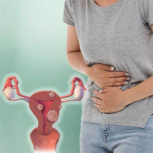
Types of Uterine Fibroids

Intramural Fibroids

Subserosal Fibroids

Submucosal Fibroids

Pedunculated Fibroids
Causes of Uterine Fibroids
Symptoms of Uterine Fibroids
The exact cause of uterine fibroids is not fully known, but several factors are believed to contribute to their development:
-
Genetic changes: Abnormal changes in genes of uterine muscle cells may lead to fibroid formation.
-
Hormones : Estrogen and progesterone promote the growth of fibroids, which is why they often reduce after menopause.
-
Growth factors: Substances like insulin-like growth factor may influence fibroid growth.
-
Extracellular matrix (ECM). ECM is the material that makes cells stick together, like mortar between bricks. ECM is increased in fibroids and makes them fibrous. ECM also stores growth factors and causes biologic changes in the cells themselves.
Many women with uterine fibroids do not experience any symptoms. When symptoms do occur, they depend on the size, number, and location of the fibroids.
Common symptoms include:
-
Heavy menstrual bleeding
-
Periods lasting longer than a week
-
Pelvic pain or pressure
-
Frequent urination
-
Difficulty emptying the bladder
-
Constipation
-
Back pain or leg pain
When to See a Doctor
-
Pelvic pain that doesn't go away
-
Overly heavy, prolonged or painful periods
-
Spotting or bleeding between periods
-
Difficulty emptying your bladder
-
Unexplained low red blood cell count (anemia)
Diagnosis of Uterine Fibroids
-
Blood test: You may be required to go for a blood test which will show your complete blood count in order to check for anemia, other blood disorders or thyroid issues.
-
Transvaginal Ultrasound: This is an imagery test that uses sound waves to produce images of the uterus. An ultrasound device called the transducer is placed inside the vagina to confirm the diagnosis and measure the fibroids.
-
Magnetic resonance imaging (MRI): This is another imagery test that can provide some detailed information regarding the size, location and type of the fibroid. Most often used in women with a larger uterus or in women approaching menopause, this test helps in determining the appropriate method of treatment.
-
Hysterosonography: Also known as saline infusion sonogram, this test uses sterile salt (saline) water to expand the uterine cavity, making it easier to get images of submucosal fibroids and the lining the uterus among women who experience heavy blood loss or in women who are attempting pregnancy.
-
Hysterosalpingography: This test uses a dye to make the uterine cavity and fallopian tubes easier to be viewed on an X-ray. Often recommended during concerns regarding infertility, this test helps the doctor to determine if your fallopian tubes are open or are blocked and can show the presence of submucosal fibroids.
-
Hysteroscopy: This test involves the insertion of a small telescope attached with a light into the uterus via the vagina and cervix. The doctor then injects saline into the uterus, expanding the uterine cavity, which allows them to examine the walls of your uterus and the openings of your fallopian tubes.
Risks & Complications During Fibroid Surgery
Fibroid removal surgery is generally safe, but like any surgical procedure, it may involve some risks depending on the patient’s condition and the type of surgery performed.
Possible complications include:
-
Allergic or adverse reaction to anesthesia
-
Heavy bleeding during or after surgery
-
Damage to the uterus or nearby organs
-
Significant blood loss that may require a blood transfusion
-
Injury to the urinary tract
Proper evaluation and experienced surgical care help minimize these risks.
How to Prevent Uterine Fibroids?



There is no sure way to completely prevent uterine fibroids, as their exact cause is still not fully known. However, certain lifestyle habits may help reduce the risk:
-
Maintain a healthy body weight
-
Eat a balanced diet rich in fruits and vegetables
-
Exercise regularly to support hormonal balance
-
Avoid excessive intake of processed and high-fat foods
-
Some studies suggest that hormonal contraceptives may be linked to a lower risk of fibroids
While prevention may not always be possible, these healthy habits can help lower the risk and support overall reproductive health.
OUR SPECIALISTS DOCTORS
Dr. ABCD
MBBS,
10 yrs of experience
Dr. ABCD
MBBS,
10 yrs of experience
Dr. ABCD
MBBS,
10 yrs of experience
Types of Surgery for Uterine Fibroids

-
Abdominal myomectomy (Open Myomectomy): This is a traditional surgical procedure in which an open incision is made in the abdomen to access the uterus and remove fibroids. After removal, the incision is closed with stitches. It is usually preferred for multiple or deeply located fibroids.
-
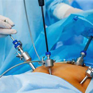
Laparoscopic myomectomy: In this minimally invasive procedure, small incisions are made in the abdomen. A laparoscope (a thin camera) is used to view the uterus, and surgical instruments are used to remove fibroids.
It results in less pain and faster recovery compared to open surgery.
-
Robotic Myomectomy : This is similar to laparoscopic surgery, but the surgeon uses a robotic system controlled from a console for more precision and control during fibroid removal.
-
Abdominal Hysterectomy : TThis is a surgical procedure to remove the uterus partially or completely, depending on the condition. In some cases, ovaries, cervix, or fallopian tubes may also be removed.
It is usually considered when other treatments fail, as it results in loss of fertility.